Research Projects
Quantitative PIV Enabled Echocardiography Techniques
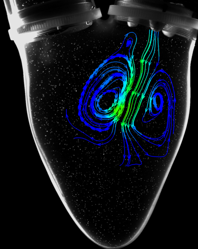
 Color Doppler imaging is a tool to obtain intracardiac flow information and considers only a single component of velocity along the transducer scan line and does not provide quantitative information with respect to vortex formation, recirculation areas, and flow stagnation zones. Because of these limitations, color Doppler imaging may not provide adequate information for either the reconstruction of velocity vector fields or the actual direction of streamlines. Quantitative piv enabled echocardiography technique is an ultrasound, non-Doppler, method to measure multi-component velocity information non-invasively. This method is the in vivo equivalent of optical particle image velocimetry (PIV) techniques and is named echo-PIV.
The echo-PIV method takes advantage of the non-linear backscatter characteristics of small gas-filled microbubbles that are introduced into the blood flow stream. Analysis of the sub- and super-harmonic signals within the backscatter allows separation of signals from tissue and flow and facilitates precise particle detection. Once particles are detected within the flow field, particle image velocimetry algorithms can be applied to determine the local velocity vector.
