
Research Projects
Artificial Intelligence (Deep-learning) in Cardiovascular Imaging
The field of artificial intelligence (AI) offers opportunities to significantly improve the speed, accuracy, and quality of image interpretation and diagnosis in radiology. AI aims to mimic human cognitive functions. Our solutions based on Artificial Neural Networks (ANNs) and machine learning concepts eliminate inter- and intra-observer discrepancies and improve clinical workflow efficiency and reproducibility.
A combined deep-learning and deformable-model approach to fully automatic segmentation of the left and right ventricles in cardiac MRISegmentation of the right and left ventricles from cardiac MRI datasets is an essential step for calculation of clinical indices such as ventricular volume, ejection fraction, left ventricular mass and wall thickness. Manual delineation by experts is currently the standard clinical practice for performing the ventricle segmentation. However, manual segmentation is tedious, time consuming and prone to intra- and inter-observer variability. In collaboration with Prof. Jafarkhani at UCI, we have been working on cutting-edge AI solutions to this problem for years. In addition, we have leveraged different state-of-the-art image processing methods including region growing, deformable models and supervised learning algorithms.
We have developed and validated a fully automated, accurate and robust segmentation method from cardiac MRI. In terms of novelty and contributions, our work is one of the early attempts of employing deep learning algorithms for cardiac MRI segmentation. The following block diagram summarizes our approach. For further information please look at our paper at Medical Image Analysis and Magnetic Resonance in Medicine. Computer codes can also be accessed at LV-segmentation-in-cardiac-MRI-master.zip and RVsegCardiacDLcombined-master.zip, respectively.
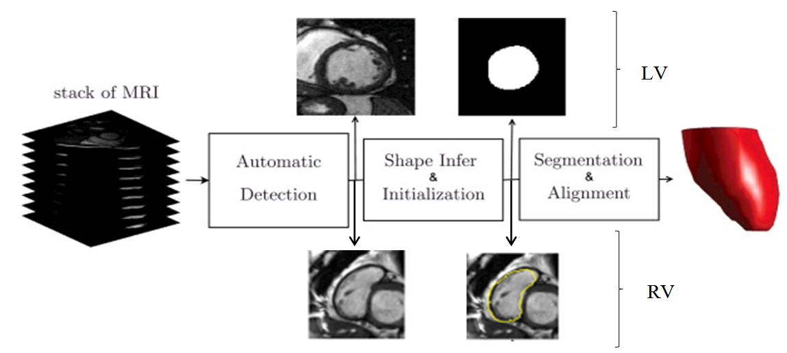 Block diagram of the integrated deep learning and deformable model algorithm for RV segmentation. The image is adapted from Avendi et al., 2016 and 2017.
A 3-D active contour method for automated segmentation of the left ventricle from magnetic resonance images
Previously, most cardiac segmentation techniques treated 2-D segmentation and 3-D multiplanar reconstruction as two separate processes. These processes achieve volumetric reconstruction by first applying a 2-D segmentation approach independently for each slice, and then volumizing these 2-D segmented image stacks into 3-D objects. To achieve the best results for 3D reconstruction, we take on a novel approach which is true 3-D reconstruction and exploits the benefit of full volumetric imaging, which does not rely on prior statistical knowledge. Our method is carried out over three consecutive steps, endocardial segmentation, intrachamber inclusion using convex-hull interpolation and myocardial segmentation. For further information please look at our journal article in IEEE Transaction of Biomedical Engineering.
View all research projects